Digital dermatoscopy
From early childhood to old age, moles constantly appear, and those that are present can change. It is not possible to completely prevent moles, it depends on genetics, sun exposure, lifestyle. Taking care of your skin should be natural for all of us.
Dermatoscopy
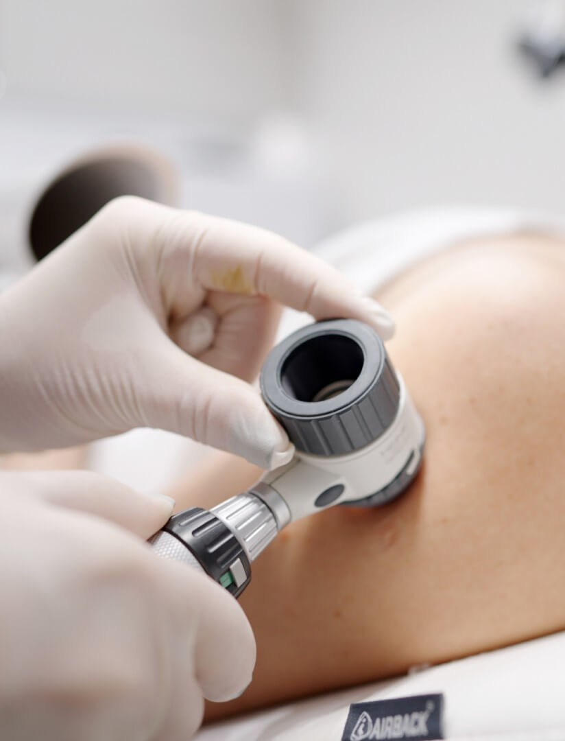
Dermatoscopy is a diagnostic tool for assessing skin changes using a dermatoscope, i.e. an instrument that magnifies the field of view with polarised or unpolarised light. Dermatoscope is a very important tool in the assessment of pigmented skin changes. It is performed after a clinical skin examination and allows the epidermis and the deeper upper dermis to be viewed.
It helps the dermatologist to distinguish between melanocytic and non-melanocytic changes and is of the greatest importance for the early diagnosis of melanoma. Melanoma is the most malignant skin cancer, which in about 50% of cases arises in healthy skin as a new formation, and in 50% form changing nevi. If diagnosed early, melanoma is a curable cancer. Unlike other cancers, it is often easy to detect through regular skin checks with a specialist.
Dermatoscopy helps to differentiate various skin tumors – both benign and malignant (keratoses, dermatofibromas, hemangiomas, warts, basaliomas, squamous cell carcinomas, melanomas, and other lesions). Dermatological examination helps to diagnose the type of formation and determine when treatment is needed.
Digital dermatoscopy
Digital dermatoscopy allows you to monitor mole changes over time, with images of the skin lesions being saved for each examination and comparing with previous ones, if significant changes are observed, our doctor will instruct you to have the lesion removed. This type of examination is particularly important for those at increased risk of melanoma: people with a large number of moles on their body, those with atypical moles; those with fair skin and high sensitivity to sun exposure (redheads with freckles); those with sunburn in childhood and adolescence; those who are exposed to UV rays on a regular basis due to a profession or a hobby; and those who have been previously treated for melanoma or other skin cancer (or a member of their family).
If you notice changes in moles such as asymmetry, irregular edges, discoloration, or an increase in the diameter of the mole, bleeding or itching, be sure to consult a dermatologist. It is recommended to test moles once a year, and for those who are at greater risk, you should visit a dermatologist more often.
At “Sapiegos Clinic”, we use the world’s most advanced digital dermatoscope, the FotoFinder, which is the gold standard in dermatology diagnostics. It is an integrated imaging system for the early detection of skin cancer, specifically designed to perform all the most important aspects of skin examination and imaging. Its accuracy makes it one of the most highly regarded diagnostic devices for skin cancer, validated by numerous scientific awards. Thanks to the synergy of the different diagnostic modules, the dermatoscope allows standardised examination of suspicious skin lesions and detailed documentation of the individual’s image, as well as continuous monitoring of moles. Using calibrated optics, the skin is magnified 20 to 140 times to ensure consistent mole analysis every time. The digital dermatoscope allows for the capture, labelling, storage, and transfer of dermatoscope images, as well as clear visualisation and analysis of suspicious lesions for the patient and the physician.

Why choose digital dermatoscopy?
- Standardised examination of moles at each regular check-up.
- Monitoring of the development of the mole over time – comparison of the mole with the previous image.
- In-depth analysis of the structure, shape, and colour of the mole.
- Artificial intelligence – to help you decide whether you need to surgically remove a mole.
Methods of mole remover
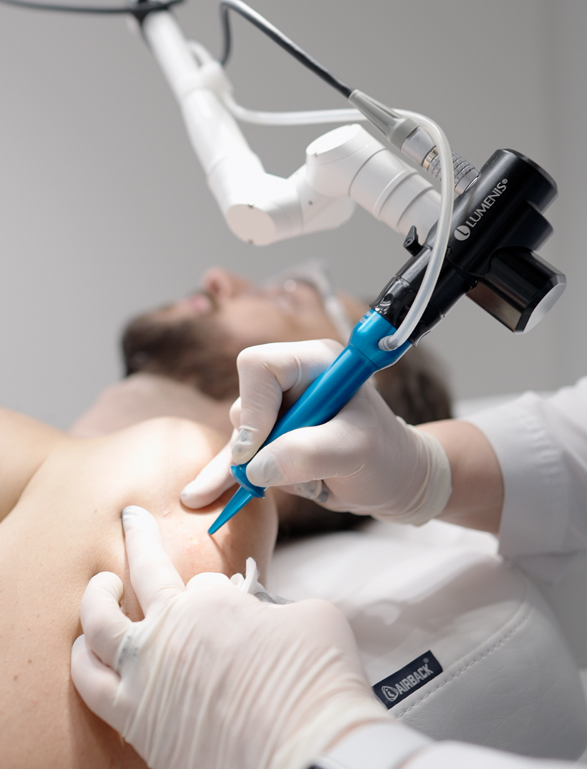
Our clinic offers two methods of removing moles and other benign lesions – laser and surgical.
The “Erbium” laser method and the CO2 laser are the simplest way to remove moles and other skin lesions. In most cases, no anaesthesia is required for removal. The laser uses a beam of light that is directed directly at the mole, heats the tissue, and then vaporises it. The procedure is painless and does not require incisions or skin sutures. Laser mole removal is less likely to leave scars. Laser treatment leaves a wound at the site of the former lesion, which heals with a scab. Healing usually takes about 1-2 weeks. It is important to note that laser treatment removes the skin at a microscopic level, so it is not possible to examine the lesion histologically to make a definitive diagnosis. Therefore, only benign lesions are removed by laser. Before removal, the lesion must be assessed by a dermatologist to ensure that it is benign.
The surgical approach involves removing the mole with a scalpel. The mole must be removed completely, i.e., the superficial part and the part under the skin. In the case of a small mole, the wound is sutured directly, but in the case of larger moles, a loose skin graft is sometimes used to cover the skin. Recovery from the procedure takes about 7-14 days. After removal of the stitches, treatment is recommended to apply the treated area with the prescribed cream for the next few weeks to soften the scar.
Prices
Dr. Dermatovenereologist consultation with dermatoscopy
99 €
FotoFinder digital dermatoscopy
39 €